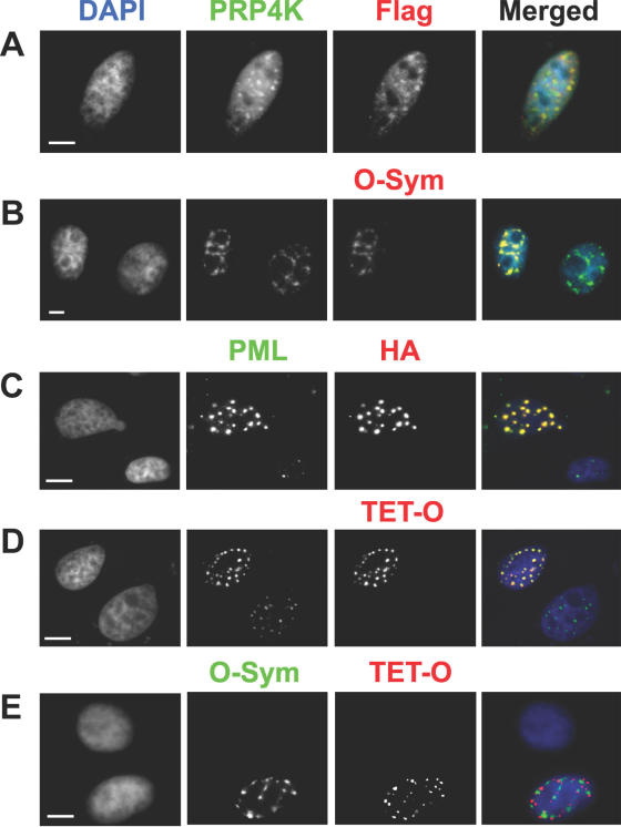
Figure 2.
In situ localization of SC35 and PML in human SK-N-SH cells using dsDNA oligonucleotides. (A) Localization of the LacI-tagged SC35 in paraformaldehyde fixed cells transfected with pGD-Flag-Lac-SC35 using the anti-Flag antibody M2 (Flag, red) or (B) with a Cy3-labelled dsDNA oligo (O-Sym, red) specific for LacI. PRP4 kinase was also localized as an endogenous marker for splicing speckles by antibody detection (PRP4K, green). Positive co-localization between PRP4K and LacI-tagged SC35 is demonstrated by a yellow signal in the merged images. The localization of the LacI–SC35 fusion protein was observed only in transfected cells (see B, O-Sym) and showed complete co-localization with PRP4 kinase (PRP4K, green) in nuclear speckles. (C) Localization of the TetR-tagged PML in paraformaldehyde fixed cells transfected with pGD-HA-TET-PML using the anti-HA (HA, red) or (D) with a Cy5-labelled dsDNA oligo (TET-O, red) specific for TetR. Endogenous PML was also localized as a marker for PML nuclear bodies antibody detection (PML, green). Positive co-localization between PML and TetR-tagged PML is demonstrated by a yellow signal in the merged images. The localization of the TetR–PML fusion protein was observed only in transfected cells (see C, TET-O) and showed complete co-localization with PML (PML, green) in PML nuclear bodies. (E) Multiple detection and localization of LacI–SC35 and TetR–PML in cells transfected with pGD-HA-TET-PML and pGD-Flag-Lac338-Sc35. Localization of LacI–SC35 and TetR–PML was accomplished using Cy3-labelled O-Sym or Cy5-labelled TET-O dsDNA oligos, respectively. TetR–PML and LacI–SC35 do not co-localize [separate red and green signals (respectively) in merged image], thus demonstrating the utility of dsDNA oligos for the multiplex detection of proteins in situ. DNA was counterstained with DAPI (blue in merged images). Scale bars = 5 μm.

