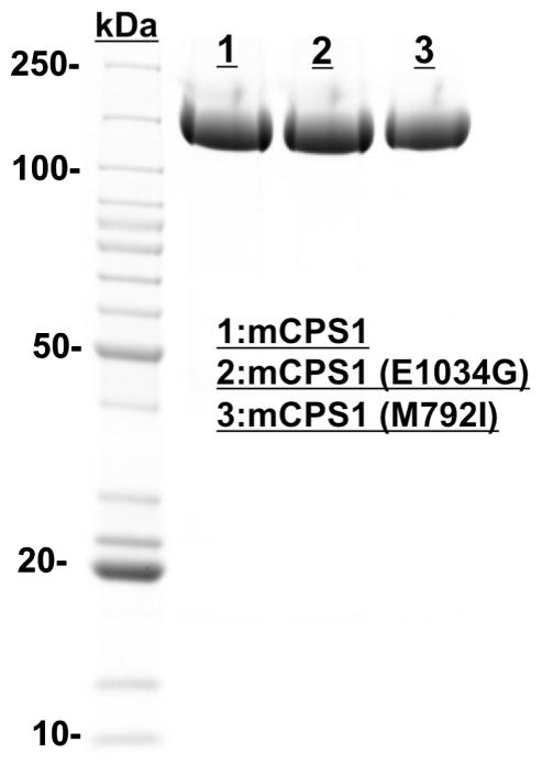
Fig. 1.
SDS/PAGE (Coomassie Blue staining, ~10 μg of total protein per lane) of purified wild-type mouse CPS1 and two mutants (E1034G and M792I) illustrating their expression levels. Lane1, recombinant wild-type mouse CPS1 (mCPS1); Lane 2, E1034G mouse CPS1 mutant; Lane 3, M792I mouse CPS1 mutant. (For interpretation of the references to colour in this figure legend, the reader is referred to the web version of this article.)

