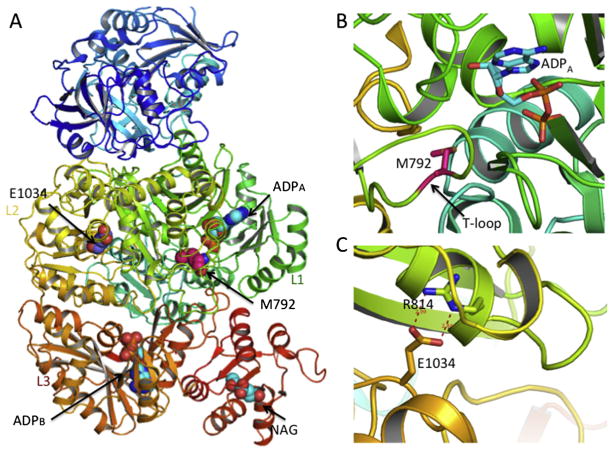
Fig. 6.
Locations of E1034G and M792I mutation in the CPS1 structure. (A) Ribbon diagram of the human CPS1 structure bound with NAG (sphere representation) and ADP showing the location of E1034 and M792. (B) Details of M792 site. The side-chain of M792 and the bound ADP are shown in sticks. (C) Details of E1034 site with hydrogen bonding interactions shown in dashed lines.

