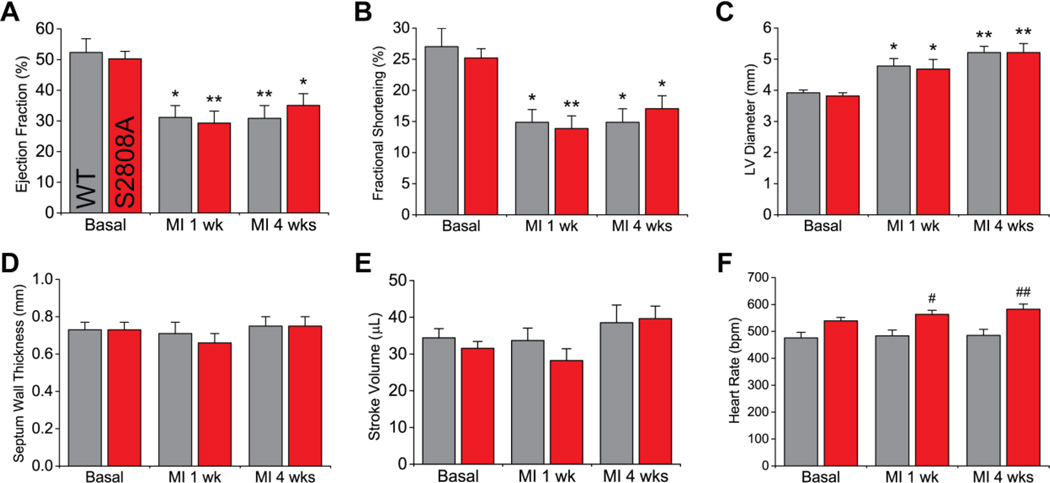
Figure 2. Cardiac function after MI.
MI due to permanent LAD ligation produced a significant decrease in ejection fraction (A) and fractional shortening (B), and an increase in LV diameter in diastole (C). Septum wall thickness in diastole (E) and stroke volume (E) remained unaltered. Most parameters, measured at one and four weeks after MI, were comparable between S2808A and WT mice, except for heart rate (F) (n = 9 per genotype; * p < 0.05, ** p < 0.01 vs. same genotype basal; # p < 0.05, ## p < 0.01 vs. WT at same time-point).

