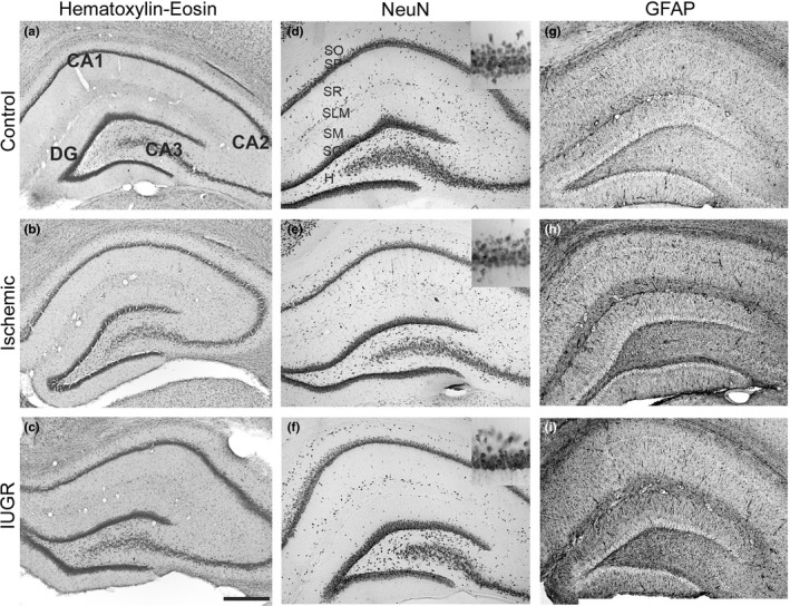
Figure 3.

Hippocampus histology. Coronal hippocampal sections of control (a, d, g,) ischemic (b, e, h) and Intrauterine growth restriction (IUGR) (c, f, i) rats at P35. (a–c) Nissl stained; d–f) immunostained against NeuN, inset correspond to a higher magnification of the CA1 pyramidal layer; (g–i) GFAP immunostained. CA1, CA2, CA3 hippocampal fields; DG dentate gyrus; H, hilus; SO, stratum oriens; SP, stratum pyramidale; SR, stratum radiatum; SG, stratum granulosum; SLM, stratum lacunosum‐moleculare; SM, stratum moleculare; Scale bar: 150 μm
