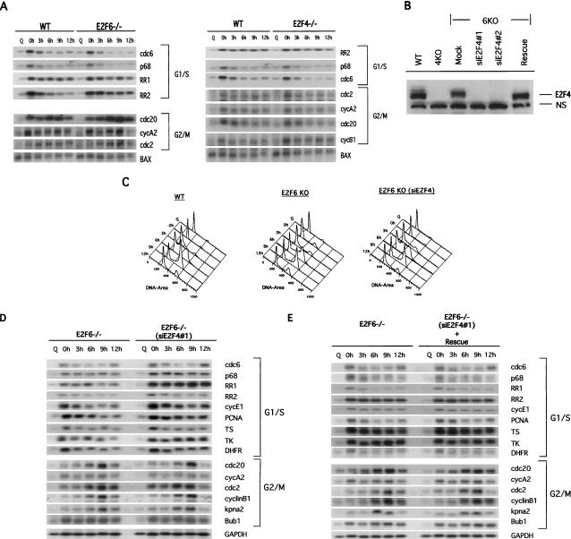
Figure 5.
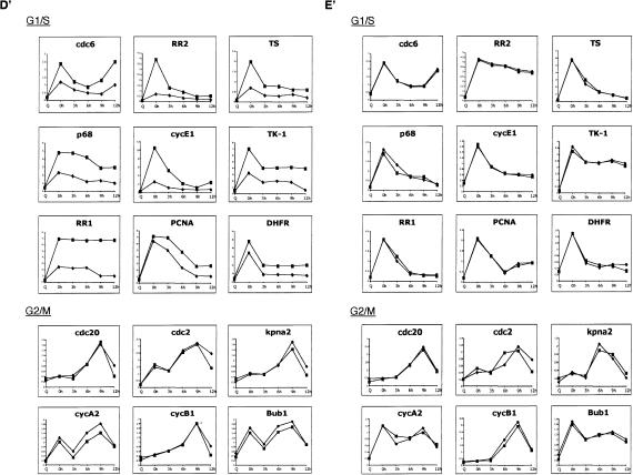
Role of E2F6 in E2F-target gene expression. (A) Wild-type (WT), E2F4-null, and E2F6-null MEFs were synchronized by serum starvation (Q, quiescence) and then stimulated by the addition of serum and 0.5 mM hydroxyurea (HU) to block cells at the G1/S boundary. Cells were then released from the HU block using media containing 15% FBS and allowed to progress through the cell cycle. Cells were harvested for RNA at 0, 3, 6, 9, and 12 h after HU release. Equal amounts of mRNA were subjected to Northern blot analysis to determine the pattern of expression of a number of E2F-responsive genes.(B) Extracts of E2F6-/- cells infected with either Retrovirus alone (Mock), Retrovirus encoding siRNAs against E2F4 (siE2F4#1 and siE2F4#2), or siE2F4#1 along with pCDNA3-E2F4m (Rescue) were resolved on SDS acrylamide gels and assessed for presence of E2F4 protein by Western blotting with specific antibodies. (N.S.) Nonspecific protein. Extracts from wild-type (WT) and E2F4-/- cells (4KO) were included as controls. (C) FACS analysis of wild-type (WT), E2F6-/- (E2F6 KO), and E2F6-/- cells infected with an siE2F4-expressing retrovirus [E2F6 KO(siE2F4)]. Cells were synchronized by a hydroxyurea (HU) block, and then released into the cell cycle. (Q) Quiescent cells. Time points were collected at 0, 3, 6, 9, and 12 h after release from HU block. (D) Northern blot analysis of E2F6-/- cells transfected with either a pool of four nonspecific siRNAs (control) or an siRNA specific to E2F4 (siE2F4#1). Cells were synchronized by serum starvation (Q, quiescence) and then stimulated by the addition of serum and 0.5 mM hydroxyurea (HU) to block cells at the G1/S boundary. Cells were then released from the HU block using media containing 15% FBS and allowed to progress through the cell cycle. Cells were harvested for RNA at 0, 3, 6, 9, and 12 h after HU release. Equal amounts of mRNA were subjected to Northern blot analysis to determine the pattern of expression of a number of E2F-responsive genes. (D′) The expression level of the genes shown in D was quantitated by PhosphorImager analysis and normalized to the GAPDH control. E2F6-/- (diamond); siE2F4#1 (square). (E) Northern blot analysis of E2F6-/- cells transfected with either a pool of four nonspecific siRNAs (control), or siE2F4 plus an expression plasmid for human E2F4 (Rescue). The human E2F4 cDNA was mutated to prevent degradation of exogenous E2F4 mRNA and therefore depletion of E2F4 protein levels. Cells were treated as above and harvested for RNA at 0, 3, 6, 9, and 12 h after HU release. Equal amounts of mRNA were subjected to Northern blot analysis to determine the pattern of expression of several E2F-responsive genes. (E′) The expression level of the genes shown in E was quantitated by PhosphorImager analysis and normalized to the GAPDH control. (Diamond) E2F6-/-; (square) Rescue.


