Figure 1.
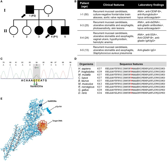
(A) Family pedigree of patients. Symbols in black indicate individuals with the same genetic defect and arrow signs indicate the patients enrolled in this study. (B) Table showing clinical data of all four affected individuals. (C) Sanger DNA sequencing chromatogram of mutated STAT1 gene. (D) Evolutionary conservation of p.Val653 among species. (E) Three-dimensional structure of phosphorylated STAT1 protein with the mutation (Val653Ile), the phosphorylation site (pTyr701), and the target DNA indicated.

