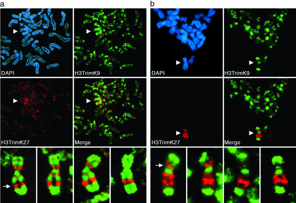
Fig. 1.
Spatial relationship of two major Xi heterochromatin types at metaphase. Images represent typical distributions obtained from three independent female cell lines. (a) Partial metaphase spread of RPE1 cells showing the spatial distribution of H3TrimK9 (green, FITC) and H3TrimK27 (red, rhodamine) and four additional higher-magnification images of the Xi showing the merged H3TrimK9 and H3TrimK27 distributions. The white arrow indicates the major H3TrimK27 band centered at Xq23. (b) Distributions of H3TrimK9 and H3TrimK27 in HME1 cells. The location of the Xi in the partial metaphase spreads is indicated by white arrowheads. The white arrow indicates the major H3TrimK27 band centered at Xp11. All images were obtained by indirect immunofluorescence.

