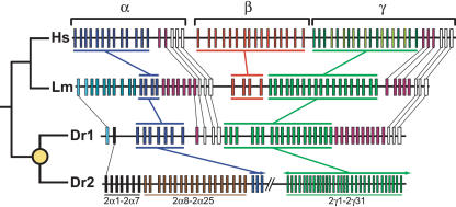
Figure 4.
Comparison of the human (Hs), coelacanth (Lm), and zebrafish (Dr1 and Dr2) protocadherin clusters. The known phylogeny is shown at left. The yellow circle indicates the teleost whole-genome duplication. Genes in each paralog subgroup are indicated by color and connecting bars. Individual orthologous relationships are indicated by narrow lines. Zebrafish-specific paralog subgroups in DrPcdh2α are labeled. Sequencing of DrPcdh2 is sufficiently complete to determine that β protocadherins are absent. DrPcdh2γ exons are numbered on the basis of their order in the genomic contig BX005294, not on their order in the genome.

