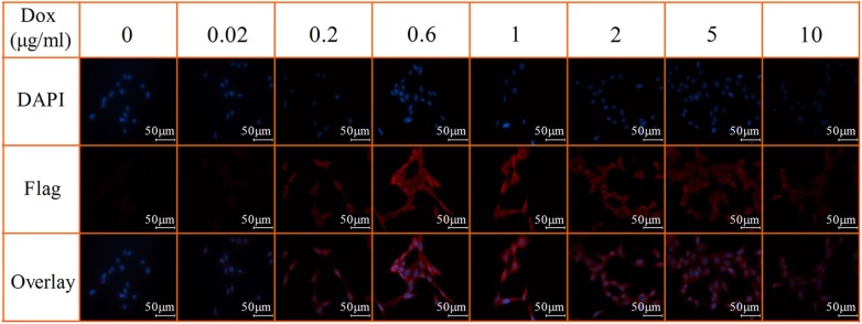
Figure 3. Immunofluorescent staining of Dox-inducible Flag expression.
SK-N-SH-FTH1 cells were cultured for 72 h in various concentrations of Dox. Immunostaining with a flag-specific antibody showed that flag expression (red fluorescence) was significantly stronger in SK-N-SH-FTH1 cells after being exposed to 0.6 μg/ml Dox for 72 h, which was consistent with the western blot results. Cell nuclei were stained with DAPI (blue). The scale bars all represent 50 μm.

