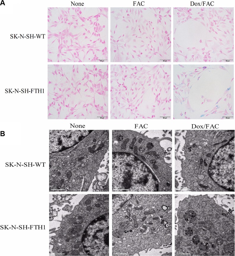
Figure 5. Intracellular iron accumulation in SK-N-SH-FTH1 and SK-N-SH-WT cells.
(A) Prussian blue staining revealed blue iron particles distributed throughout the cytoplasm of SK-N-SH-FTH1 cells but few particles dispersed within SK-N-SH-WT cells upon Dox induction and FAC supplementation (Dox/FAC, 0.6 μg/ml Dox and 500 μM FAC). Few blue particles were present within the SK-N-SH-WT and SK-N-SH-FTH1 cells treated only with FAC (FAC group). No blue particles were detected in SK-N-SH-FTH1 or SK-N-SH-WT cells in the absence of both Dox and FAC (None group). (B) The TEM results, which showed black iron particles accumulated in cytoplasmic vacuoles, were consistent with those of Prussian blue staining. The scale bars represent 50 μm (A) and 1 μm (B).

