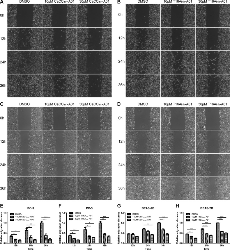
Figure 7. Suppression of cell migration of PC-3 and BEAS-2B cells by ANO1 inhibition in wound-healing assay.
Cell migration was assessed by wound-healing assay in the presence of ANO1 inhibitor or DMSO control. Images of wound closure in prostate cancer PC-3 cells treated with different concentrations of CaCCinh-A01 (A) or T16Ainh-A01 (B) were taken at 0 h, 12 h, 24 h and 36 h. Representative images of bronchial epithelial BEAS-2B cells treated with CaCCinh-A01 (C) or T16Ainh-A01 (D) were presented. Scale bar: 100 μm. (E–H) Bar graphs of panel A–D showing treatment of PC-3 and BEAS-2B cells with CaCCinh-A01 or T16Ainh-A01 suppressed cell migration. Data are expressed as mean ± SEM; n = 3; * P < 0.05; ** P < 0.01;*** P < 0.001.

