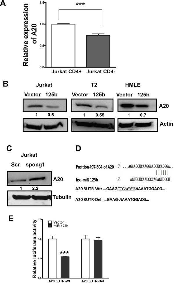
Figure 2. A20 is a direct target of miR-125b in T cells.

A. qRT-PCR was performed to examine the expression of A20 in CD4+population and CD4-pouplation of Jurkat cell lines. GAPDH was used as an internal control and for normalization of the data. Columns represent the mean of three independent experiments; bars represent SE. ***, p<0.001. B and C. Jurkat-vector, Jurkat-miR-125b, T2-vector, T2-miR-125b, HMLE-vector, HMLE-miR-125, Jurkat-scramble and Jurkat-miR-125b-spong1 stable cell lines were collected. Cell lysates were prepared for Western blotting with an antibody against A20 with β-actin used as a loading control. D. A20 3′ UTR contains a predicted miR-125b-binding site. Alignment between the miR-125b seed sequence and A20 3′ UTR is shown. A schematic diagram shows the wild type and mutant 3′UTR of A20. E. Jurkat cells were co-transfected with luciferase reporter plasmids with or without 3′-UTR of A20, pre-miR-125, or pre-miR-negative (Ctr). 48 hours post-transfection, cells were harvested and lysed with lysis buffer. Luciferase activity was measured by using a dual luciferase reporter assay. The pRL-TK vector was used as an internal control. The results were presented as relative luciferase activity (firefly LUC/Renilla LUC).
