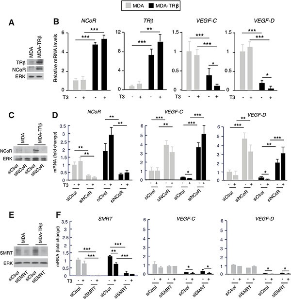
Figure 2. NCoR depletion increases VEGF-C and VEGF-D gene expression.

(A) Western blot analysis of TRβ and NCoR in parental MDA-MB-231 cells and in cells stably expressing the receptor (MDA and MDA-TRβ, respectively). ERK was used as a loading control. (B) mRNA levels of the indicated genes were determined in cells treated in the presence and absence of 5 nM T3 for 36 h. (C) NCoR and ERK levels after 72 h of transfection with siControl or siNCoR. (D) Transcript levels of NCoR, VEGF-C and VEGF-D in cells transfected with siControl or siNCoR and treated with and without T3. (E) SMRT and ERK levels after 72 h of transfection with siControl or siSMRT. (F) Transcript levels of SMRT, VEGF-C and VEGF-D in cells transfected with siControl or siSMRT and treated with and without T3. All data are means ± S.D and are expressed relative to the values obtained in untreated parental cells transfected with the control siRNA. Significance of ANOVA post-test among the indicated groups is shown as * P < 0.05, **P < 0.01 and ***P < 0.001.
