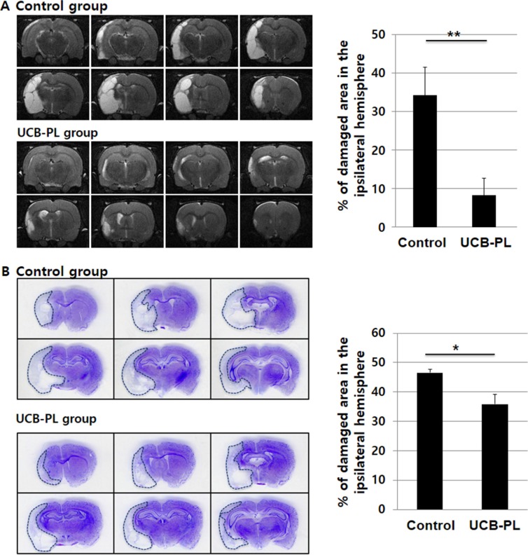
Figure 3. UCB-PL administration reduced brain tissue damage caused by MCAO injury.
(A) (Left panel) In vivo brain MRI was performed prior to euthanasia to measure the size of the damaged brain area. (Right panel) The percentage of the damaged area in the ipsilateral hemisphere was compared between the control and UCB-PL groups. Eight sections per rat were analyzed to quantify the percentage of the remaining intact ipsilateral area. (B) (Left panel) Cresyl violet staining of the rat brains was performed to measure the size of the damaged brain area. (Right panel) The graph indicates the percentages of the hemispheres that were damaged by ischemia in each group. n = 6/group, *p < 0.05, **p < 0.01.

