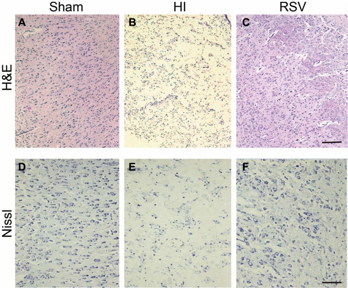
Figure 2. General morphology of the cortex after hypoxic-ischemia.
(A–C) Representative microphotographs of hematoxylin and eosin (H&E) stained cortex from (A) Sham, (B) HI and (C) RSV at 7-day after hypoxic-ischemic insult. Scale bar = 200 μm. (D–F) Representative photomicrographs of Nissl stained cortex from (D) Sham, (E) HI and (F) RSV animals at 7-day after the injury. Scale bar = 100 μm.

