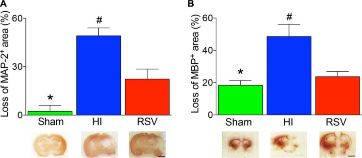
Figure 3. Lesion size with MAP-2 and MBP staining after hypoxic-ischemic injury.
The loss of MAP-2 (A) and MBP (B) areas were significantly higher in the HI group than in the groups of Sham or RSV. Representative examples of coronal sections at 7 day after hypoxic-ischemia were shown in the lower panels accordingly. Values are mean ± SEM, n = 4 animals per group. Sham vs. RSV, *p < 0.05; HI vs. Sham or RSV, #p < 0.05.

