
Figure 5. Bright-field pictures.
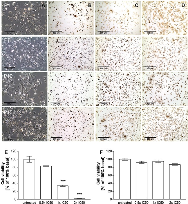
A. as well as Ki67 B., SF-1 C. and 3βHSD D. stainings of MUC-1 cells in passages 4, 7, 10 and 13 in vitro. Treatment dependent inhibition of cell viability of NCI-H295R E. and MUC-1 F. upon addition of different concentrations of EDP-M in vitro. Stars represent statistical significance over untreated controls.