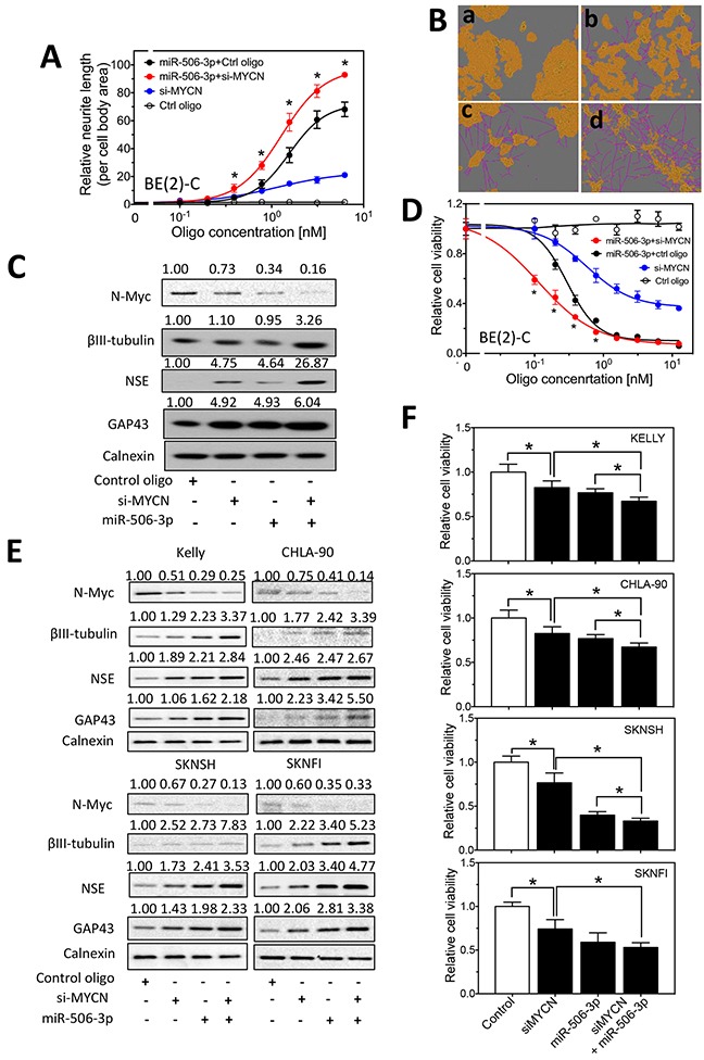
Figure 5. Effect of MYCN knockdown and miR-506-3p overexpression on cell differentiation and cell survival in neuroblastoma cells.

A-B. Effect of MYCN knockdown and miR-506-3p overexpression on neurite outgrowth in BE(2)-C cells. Cells were transfected with different concentrations of the indicated oligos. After 4 days, relative neurite outgrowth was measured. (A) Quantification of neurites showing the dose-dependent effect of MYCN siRNA (si-MYCN) and miR-506-3p mimic on neurite outgrowth. (B) Representative images, with the cell body area (yellow) and neurites (pink) defined, showing the effect of si-MYCN and miR-506-3p mimic on neurite outgrowth. Shown are cell images for (a) control oligo (6 nM), (b) si-MYCN (3 nM), (c) miR-506-3p mimic (3 nM) and (d) si-MYCN (3 nM)+miR-506-3p mimic (3 nM). C. Effect of MYCN knockdown and miR-506-3p overexpression on the expression of N-Myc and differentiation markers in BE(2)-C cells. Cells were transfected with the indicated oligos. After 4 days, cell lysates were collected, and protein levels of N-Myc, as well as protein levels of the differentiation markers, βIII–tubulin, NSE and GAP43 were measured by Western blots with calnexin levels as a loading control. D. Effect of MYCN knockdown and miR-506-3p overexpression on cell viability of BE(2)-C cells. Cells were transfected with different concentrations of the indicated oligos. After 4 days, cell viability were measured as described in the Material and Methods. *, p<0.05 compared cells co-transfected with si-MYCN and miR-506-3p mimic to cells co-transfected with control oligo and miR-506-3p mimic. E. Effect of MYCN knockdown and miR-506-3p overexpression on expression of N-Myc and differentiation markers in KELLY, CHLA-90, SKNSH and SKNFI cells. F. Effect of MYCN knockdown and miR-506-3p overexpression on cell viability in KELLY, CHLA-90, SKNSH and SKNFI cells. *, p<0.05.
