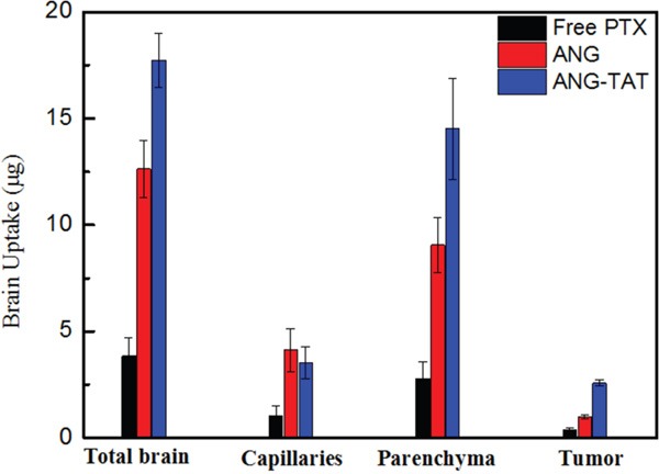
Figure 3. Brain uptake of ANG-TAT measured by in situ brain perfusion.

CD-1 mice were perfused with biotin labeled ANG-TAT (50 μM) for 5 min. After perfusion, brain capillary depletion was performed on the mice right brain hemispheres. The amount of labeled biotin associated with total brain homogenate, the brain capillary fraction and with the parenchyma was evaluated.
