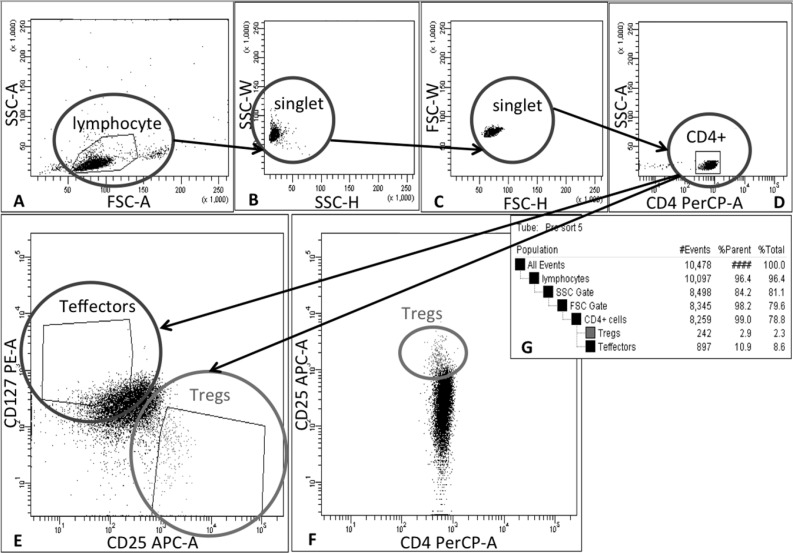
Figure 4. Schema of gating strategy for Treg sorting.
Tregs were isolated in 2-step process: firstly, CD4+ cells were pre-enriched from leukapheresis product via immunomagnetic positive selection on CliniMACS® device and then cells were stained with monoclonal antibodies and separated by Fluorescence Activated Cell Sorting (FACS). During FACS cells were gated as follows: (A) lymphocytes were identified on forward (FSC) and side-scatter (SSC) plot, (B) and (C) doublets were excluded by applying SSC-H vs SSC-W and FSC-H vs FSC-W gates, (D) followed by CD4+ cell gating. (E) Finally, Tregs and Teffectors were gated as CD25hiCD127lo/− and CD25lo/−CD127+ cells, respectively; (F) dot-plot CD4 PerCP vs CD25 APC was used as a reference for correct Treg gating based on lower CD4 expression in Treg population than in Teffectors. (G) table with population hierarchy and statistics generated by FACSDiva Software during cell sorting on FACSAria III cell sorter (BD Biosciences, San Jose, CA, USA

