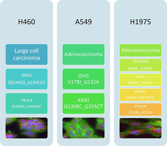
Figure 2. Histological and mutational characterisation of NSCLC cell lines.

NSCLC cell lines used in this study were chosen based on their differing histological subtypes. Mutation status was obtained from the COSMIC database. Cell images were obtained using the InCell 1000. Each cell line was stained with Höechst blue nuclear stain, mitotracker red mitochondrial stain and phalloidin green F-actin stain.
