
Figure 3. Epithelial cancer cells with co-expression of BRGs induced by CAF/BMP2 signaling gain the advantages of homing to, residing in and growing in an osteoblast-mimic bone microenvironment.
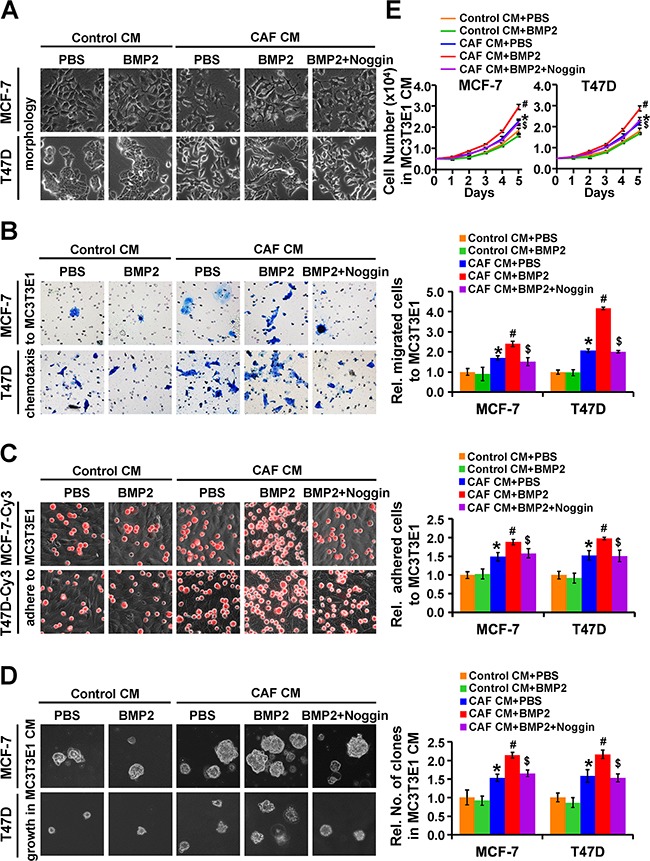
MCF-7 and T47D cells were treated as indicated. A. The morphology of the cancer cells. B. The chemotactic migration of cancer cells towards MC3T3E1 cells was assessed by transwell assay. C. The adhesion of cancer cells (red, labeled with Cy3) to MC3T3E1 cells was assessed by putting cancer cells on top of MC3T3E1 cells at 100% confluence and incubating the co-culture for 30 min. D. The colony formation of cancer cells in soft agar containing MC3T3E1 CM. Magnification: 200X. E. The proliferation of cancer cells in MC3T3E1 CM. Data are presented as the mean ± S.D. of three independent experiments performed in duplicate. *, P < 0.05 compared with Control CM plus PBS; #, P < 0.05 compared with cells treated with CAF CM plus PBS; $, P < 0.05 compared with cells treated with CAF CM plus BMP2.