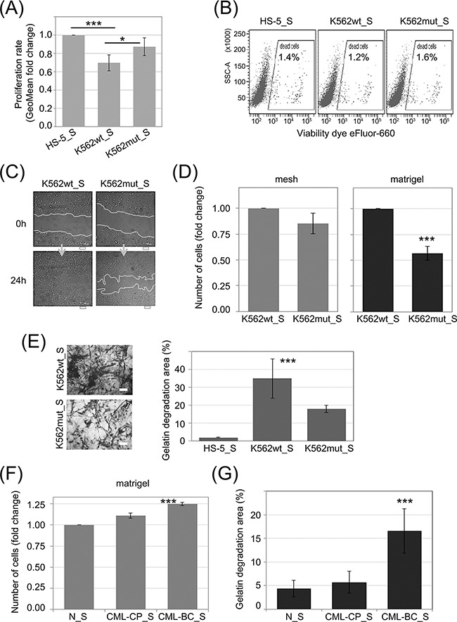
Figure 3. Influence of eIF2α phosphorylation on the invasion potential of HS-5 stromal fibroblasts.

HS-5 cells were cultured in conditioned media cleared by multistep centrifugation (K562wt_S, K562mut_S or HS-5_S). A. Proliferation rate was assayed by flow cytometry. Bars correspond to mean fold change of proliferation dye fluorescence (GeoMean) at 24h and 72h ± SEM; n=4 B. Flow cytometry analysis of cell death levels in HS-5 cells cultured in the presence of conditioned media from different cultures. Typical scatterplots for each culture condition are shown, with side scatter area (SSC-A; y-axis) plotted against viability dye eFluor 660 signal intensity. Gate used to detect dead cells is indicated on each plot C. Monolayers of HS-5 cells cultured in the presence of K562wt_S or K562mut_S were subjected to the wound healing assay. Representative images out of 3 independent experiments of wound closure immediately after wounding (0h) and after 24h are shown. Scale bar = 100 μm D. HS-5 cells were subjected to the transwell invasion assay as in Figure 2B, n=3 E. Gelatin degradation by HS-5 cells cultured in S medium, as indicated, was quantified using OregonGreen-labeled gelatin. Representative images of fluorescent gelatin (grey) are shown in the left panel. Gelatin degradation is apparent as black areas on a grey background. Scale bar = 10 μm. Right panel shows mean percentage of gelatin degradation area normalized to cell number ± SEM; n=3 independent experiments F, G. Matrigel invasion (F) and gelatin degradation (G) assays were performed as in D. and E., respectively, on HS-5 cells cultured in media conditioned by Lin-CD34+ cells from a healthy donor (N_S), chronic phase CML (CML-CP_S) or blast crisis (CML-BC_S). ***p<0.001 in Student's t-test.
