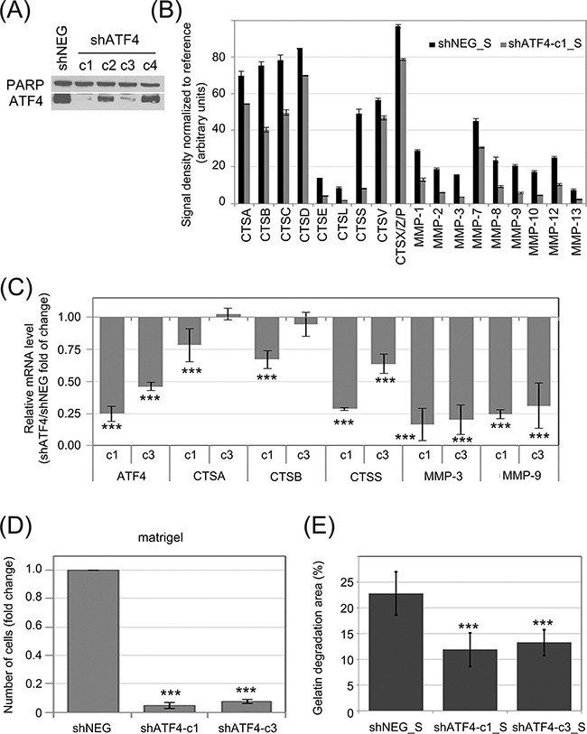
Figure 5. Influence of ATF4 on protease secretion and invasive potential of CML cells and stromal fibroblasts.

ATF4 was depleted in K562 cells by viral transduction of 4 different clones of shRNA specific to ATF4 (shATF4-c1 – c4); Non-targeting shRNA (shNEG) was used as control A. ATF4 protein level was quantified in whole cell lysates by immunoblot; PARP was used as loading control. B. Protease abundance was measured by antibody array in serum-free conditioned media from cultures of K562 expressing shATF4 (shATF4-c1_S) and control shRNA (shNEG_S). Signal density was analyzed using Image J software and normalized to reference. Graphs are mean protein level ± SEM for n=3 independent samples C. Level of mRNA of selected enzymes was measured by real-time RT-PCR and is shown as mean fold change between K562 cells expressing shATF4-c1 or shATF4-c3 and shNEG ± SEM for n=3 independent samples D. Matrigel invasion of K562 expressing the indicated shRNA constructs was measured using the transwell assay, as in Fig. 2B; n=3 independent experiments E. Gelatin degradation by HS-5 cells cultured in shATF4-c1_S or shATF4-c3_S and shNEG_S was measured as in Fig. 3E; n=3. ***p<0.001, *p<0.05 in Student's t-test.
