
Figure 4. GPC5 inhibits Wnt/β-catenin signal pathway via competitively binding to Wnt3a.
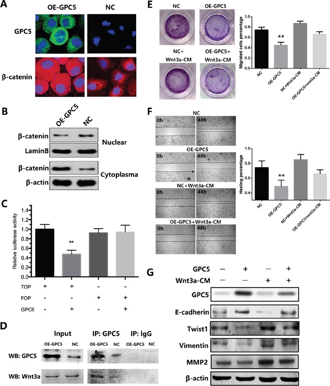
A. immunofluorescence staining of GPC5 (FITC, green, magnification: ×600) and β-catenin (TRITC, red, magnification: ×600) in OE-GPC5 A549 cells (left) and in respective control cells (right). B. the expression levels of β-catenin in nuclear and cytoplasma were compared by western blotting analysis between the OE-GPC5 A549 cells and the control cells. LaminB and β-actin were loading control respectively. C. Wnt signaling in OE-GPC5 A549 cells was detected by the TOP/FOP flash Wnt reporter assay D. in Co-immunoprecipitation assay, GPC5 was used to pull-down Wnt3a both in OE-GPC5 A549 cells and the control. E. rescue of migration by adding Wnt3a in cell culture medium of OE-GPC5 A549 cells. Representative images of transwell membrane insert in 24-well plate (left). The results were plotted as the percentage of migrated cells (right). F. representative images of Wound healing assay by adding Wnt3a in cell culture medium of OE-GPC5 A549 cells (left). And the results were plotted as the percentage of healing (right). G. the expression levels of GPC5, E-cadherin, Twist1, Vimentin and MMP2 were compared by the western blotting analysis between 4 groups in this rescue experiment.