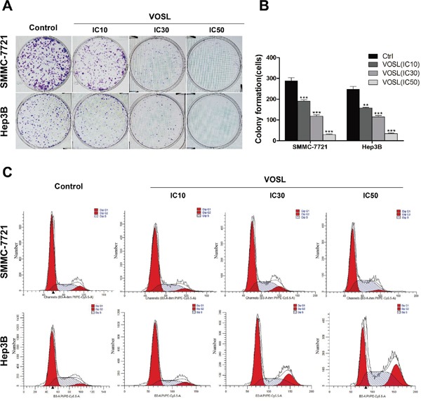
Figure 2. VOSL inhibits colony formation of HCC and influences the distribution of cell cycle.

A and B. Cells were treated with VOSL at different concentrations (IC10, IC30, IC50) for 48 h, and the colony formation was presented as mean ± SD; **p < 0.01 and ***p < 0.001 compared with the control group. C. HCC cells were exposed to VOSL (IC10, IC30, IC50) for 48 h and cell cycle was detected by flow cytometry (Detail data shown in Table 1).
