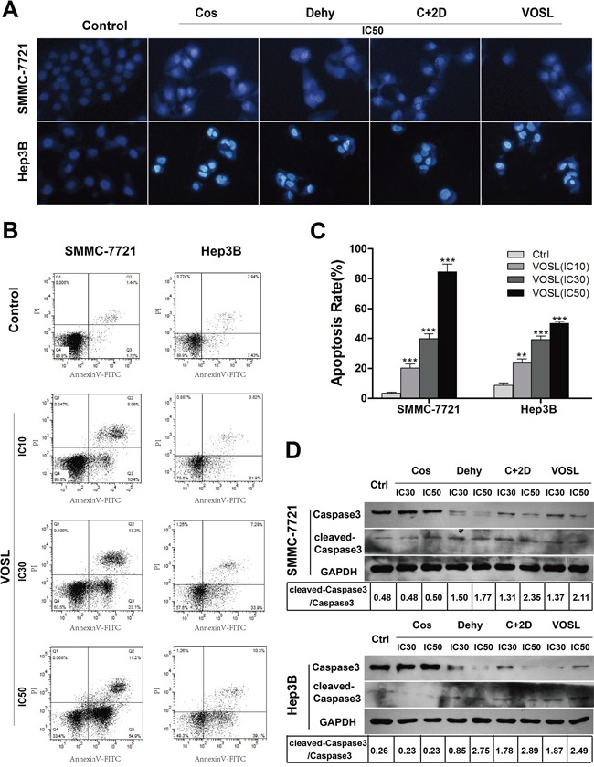
Figure 3. VOSL induces HCC cell apoptosis.

A. Apoptotic changes induced by IC50 of Cos, Dehy, C+2D snd VOSL treatments for 48 h were observed by Hoechst 33342 staining in HCC cells. B and C. Apoptotic cells were determined by flow cytometry. The apoptotic percentages were presented as mean ± SD; **p < 0.01 and ***p < 0.001 compared with the control group. D. The Caspase3 and cleaved-Caspase3 levels in HCC cells were examined by Western blot after 48 h treatment.
