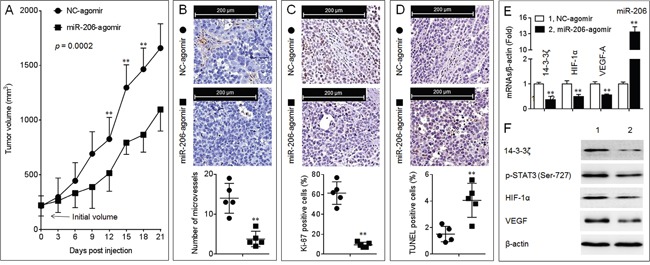
Figure 6. Effects of miR-206 on the growth, angiogenesis, and 14-3-3ζ/STAT3/HIF-1α/VEGF signal pathway in A549 xenografts tumors.

A. The volumes of xenografts tumors injected with NC-agomir or miR-206-agomir. (B., top) IHC staining of CD31. (B, bottom) The number of microvessels was quantitated based on the CD31 staining. (C., top) IHC staining of Ki-67. (C, bottom) The percentage of Ki-67 positive cells. (D, top) TUNEL staining. (D, bottom) The percentage of TUNEL positive cells. Note: Each point in (B-D) represented the mean of one xenografts tumor section calculating in 10 high-power fields. E. qPCR analyses in triplicate of the expressions of miR-206, and 14-3-3ζ, HIF-1α, VEGF-A mRNAs in A549 xenografts tumors. F. Western blots analyses of the expressions of 14-3-3ζ, p-STAT3 (Ser-727), HIF-1α, and VEGF in A549 xenografts tumors. ** p < 0.01 compared with xenografts tumors injected with NC-agomir.
