
Figure 4. RSK2 activity reduces cell adhesion to fibronectin and invasion into brain slices.
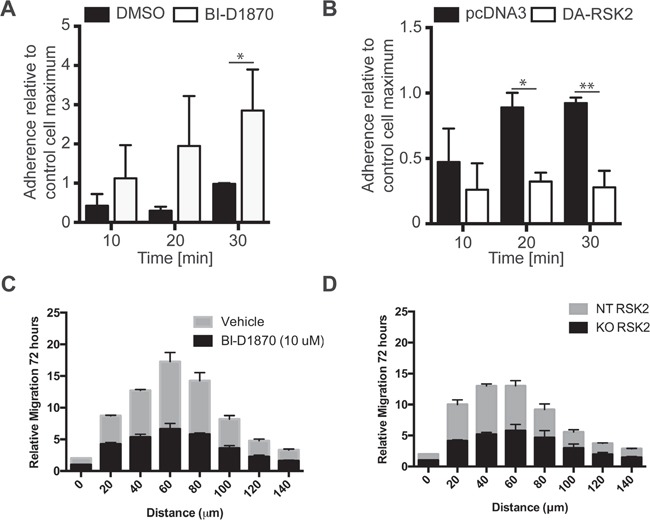
We examined early adhesion of U-87 MG cells on culture plates coated with 3FN-(9–11). Before plating, cells were A. pre-treated with 10 μM BI-D1870 or DMSO carrier control or B. transiently transfected with DA-RSK2 or a pcDNA3 empty vector control. 48 hours after transfection or 15 min after pre-treatment cell adhesion was measured. Bar graphs show the number of cells adherent after the indicated amount of time. The experiment was carried out in 3 independent repeats. Error bars shown as SEM. Statistically significant differences are marked with asterisks (* P < 0.05, ** P < 0.01, *** P < 0.001). Scale bar: 500 μm. C and D. We examined the ability of U373 cells treated with BI-D1870 inhibitor (C) or with RSK2 knocked out using CRISPR/Cas9 (D). Whole brain slices of 300 μm thickness were placed on the membrane of a six-well plate culture insert. GBM cell lines U373MG, U373MG with RSK2 gene knock-down (KO-RSK2), or U373MG with a non-targeting shRNA (NT-RSK2) were labeled with the PKH67 fluorescent linker. After 7 days, one small spheroid of approximately 200 μm was transferred to each brain slice as close to the corpus callosum as possible. The co-cultures were maintained for an additional 72 hours. To quantify the invasiveness of the spheroids, the density of the fluorescent signal was measured in each 20 μm section using ImageJ Software. Shown are the migration depths. Error bars are shown as SEM.