
Figure 2. FDG reduces Stx binding and endocytosis, and leads to increased release of the toxin back to the medium.
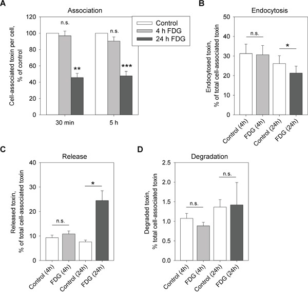
Cells were treated with 1 mM FDG for 4 or 24 h. A. 125I-Stx1-mut was added and the incubation was continued for 30 min or 5 h. Cell-associated toxin was measured and normalized to cell number. B. Cells were incubated with 125I-Stx1-mut-biotin for 20 min, the endocytosed 125I-Stx1-mut-biotin was quantified in cell lysates and normalized to the total cell-associated toxin. C and D. Cells were incubated with 125I-Stx1-mut for 30 min, the non-bound toxin was washed away and the cells were incubated with fresh medium for 1 h. The released and degraded toxin was determined as described in Materials and Methods. (C) Shows released and (D) shows degraded 125I-Stx1-mut as a percentage of total cell-associated toxin. All figures show mean values + SEM from at least three independent experiments; one-sample Student's t-test was used for (A) and paired Student's t-test was used for (B-D), *p<0.05, **p<0.005, ***p<0.0005.