
Figure 1. CD99 engagement by 0662 monoclonal antibody induces macropinocytosis in EWS cells.
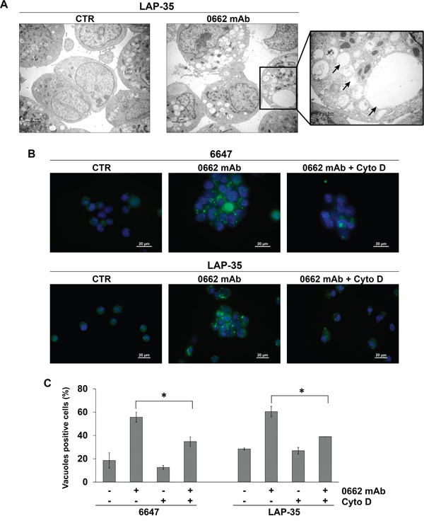
A. Transmission electron microscopy of untreated (CTR) or 0662mAb-treated LAP-35 cells (scale bar 2μm). The right panel is a magnification of mAb-treated sample (scale bar 0.5μm). Arrows indicate empty vacuoles, void of cytoplasm and organelles. B. Lucifer yellow (LY) accumulation in 6647 or LAP-35 EWS cells in presence or not of Cytochalasin D (Cyto D) after 30 min 0662mAb exposure. Images were acquired with a Nikon ECLIPSE 90i with Plan Apo 60x/NA 1.4 DIC N2 (scale bar 20μm). C. Percentage of LY-positive 6647 and LAP-35 cells in presence or absence of 0662mAb (30 min) and/or Cyto D (60 min pretreatment). Results are represented as mean ± SEM of three independent experiments (*p<0.05, Student's t test).