
Figure 9. RAS and CD99 levels are crucial for 0662mAb-induced vacuolization.
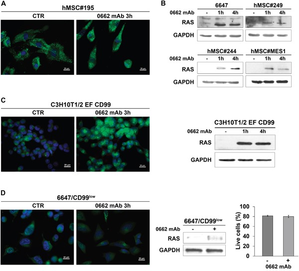
A. AO staining of human mesenchymal stem cells (hMSCs) treated or not (CTR) with the anti-CD99 0662mAb. Representative merged images are shown (scale bar 20μm). B. Western blotting evaluation of pan-RAS levels before (−) and after treatment with anti-CD99 0662mAb in 6647 cells or hMSC cells. GAPDH was used as loading control. C. Left: AO staining of murine MSCs transfected with EWS/FLI1 and CD99 (C3H10T1/2 EF CD99) treated or not (CTR) with the anti-CD99 0662mAb. Representative merged images are shown (scale bar 20μm). Right: western blotting evaluation of pan-RAS levels before (−) and after treatment with 0662mAb in C3H10T1/2 EF CD99. GAPDH was used as loading control. D. Left: AO staining of 6647/CD99low cells treated or not (CTR) with the anti-CD99 0662mAb. Representative merged images are shown (scale bar 20μm). Middle: western blotting evaluation of pan-RAS levels before (−) and after (+) treatment with 0662mAb for 1h in 6647/CD99low. GAPDH was used as loading control. Right: percentage of live cells before (−) and after (+) treatment with anti-0662mAb in 6647/CD99low (Annexin V/PI assay). Values represent mean ± SEM of three independent experiments (Student's t test: p> 0.05).