
Figure 5. Kaplan-Meier analysis of PD-L1 DNA methylation in combination with PD-L1 protein expression in prostate cancer patients (validation cohort).
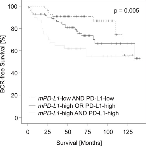
Patient samples (n = 209) were stratified according to mPD-L1 status (high vs. low) in combination with dichotomized PD-L1 protein expression status as determined earlier [5]. Patients with high PD-L1 protein expression and high mPD-L1 had shorter BCR-free intervals compared to patients with simultaneous low PD-L1 protein expression and low mPD-L1. Patients with either high PD-L1 protein expression and low mPD-L1 or low PD-L1 protein expression and high mPD-L1 showed intermediate BCR-free intervals.