Fig. 5.

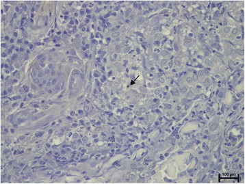
Note only one intracellular Leishmania amastigote (arrow) in the center of a granuloma in the inflammatory infiltrate present in the dermis of clinically-lesioned skin from the same dog as in Fig. 3 (Leishmania-specific IHC staining)

Note only one intracellular Leishmania amastigote (arrow) in the center of a granuloma in the inflammatory infiltrate present in the dermis of clinically-lesioned skin from the same dog as in Fig. 3 (Leishmania-specific IHC staining)