
Figure 2.
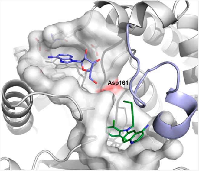
X-ray crystal structure of Dot1L with 1 and adenosine (pdb code 5mvs). Cartoon and surface representation of Dot1L (gray), flexible loop of Dot1L (126–140) (light blue), stick model of ligands 1 (green), and adenosine (blue). Asp161 is shown as red surface for orientation. The flexible loop has been omitted from the surface representation for clarity.