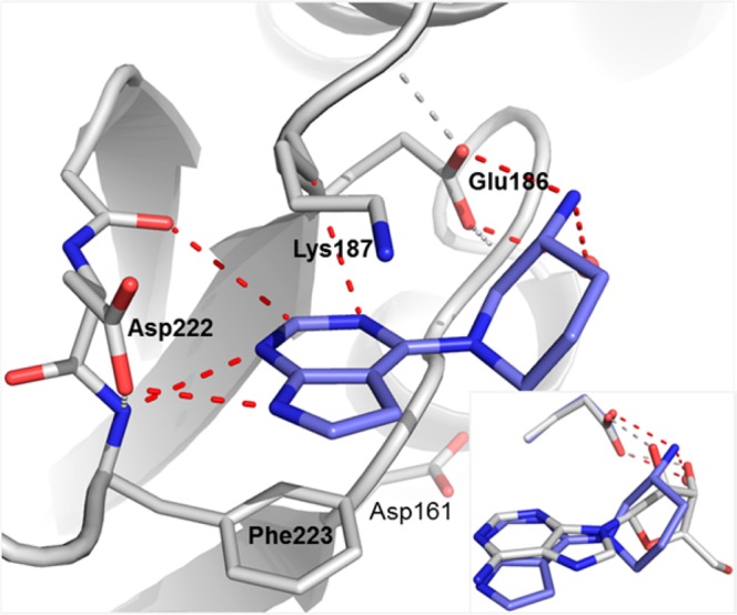
Figure 5.

Detailed interactions of 2 with Dot1L from a ternary X-ray crystal structure of Dot1L (gray) with 2 (blue) and 1 (not displayed) (pdb code 5mw3). Amino acid side chains engaging in key interactions with the ligand illustrated as sticks and polar contacts highlighted as dotted red lines (protein hydrogen bonds in gray). Inset: Overlay of adenosine (gray) and 2 (blue) based on the ternary structures. Only subtle conformational changes were observed on the protein side between the two ternary complexes.
