
Figure 4. Hyaluronan expression on cortical neurons in vitro.
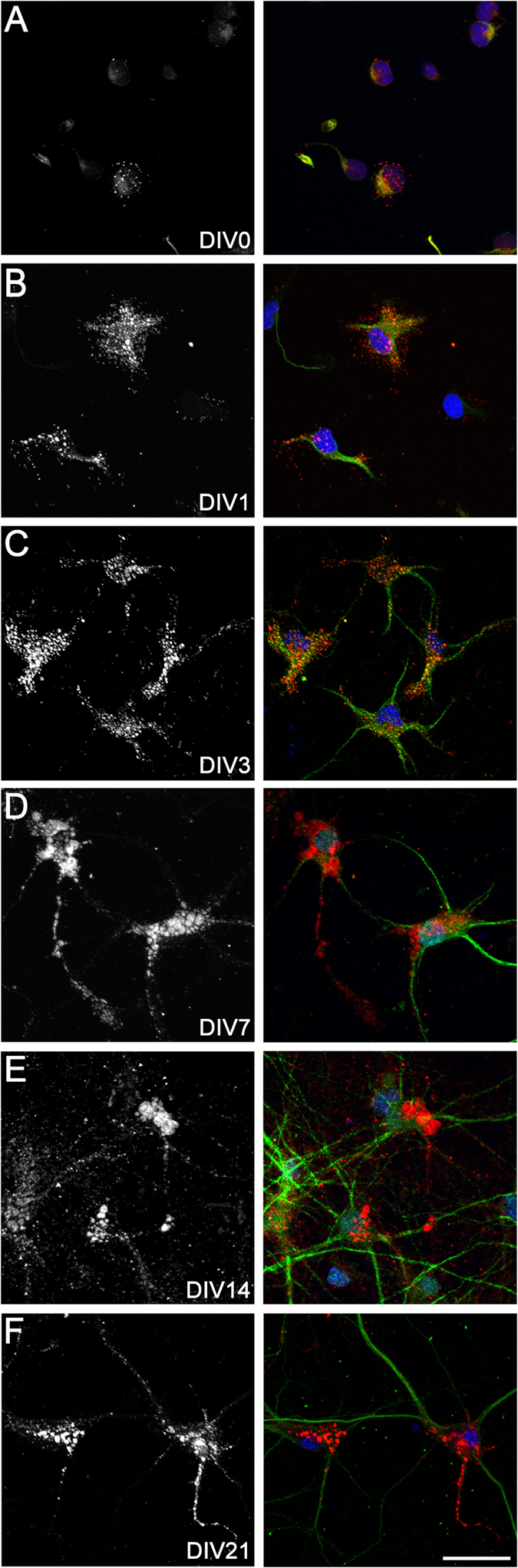
Neurons at DIV0, 1, 3, 7, 14, and 21 (A–F) were stained with biotinylated hyaluronic acid binding protein (bHABP) (left: grey, right: red), MAP2 (green), and Hoechst 33258 (blue). Scale bar: 30 μm.