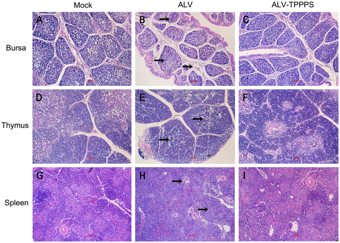
Figure 6. TPPPS treatment relieves the damage in immune organs.
Chickens in groups ALV–TPPPS and ALV were orally administered for 2 weeks with TPPPS and PBS, respectively. Non-infected chickens treated with PBS served as group Mock. Histopathological examinations were performed on the thymus, bursa, and spleen from the congenitally infected chickens at 14 dph (H&E staining, 200×). (A, D, and G) Mock-infected bursa, thymus, and spleen showed a normal morphology. (B) ALV-J-infected bursa showed lymphoid follicular dysplasia, interstitial expansion, reduced lymphocytes, and loose lymphocyte arrangement in medulla area (black arrows). (C) TPPPS-treated bursa showed an almost normal morphology. (E) ALV-J-infected thymus showed thymic hypoplasia, reduced lymphocytes, and loose lymphocyte arrangement (black arrows). (F) TPPPS-treated thymus showed minor lymphoid depletion. (H) ALV-J-infected spleen displayed hypoplasia and obvious white pulp structure (black arrows). (I) TPPPS-treated spleen showed a normal morphology. The images shown represent three animals from three independent experiments. Scale bar, 100 μm.

