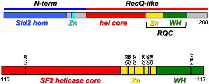
Figure 2. A schematic diagram of the domain organization of human RecQ4, showing the N-terminal region (blue) followed by helicase core (red) and the putative RQC domain (consisting of a Zn-binding domain, in yellow, and a winged helix domain, in green).
In the lower panel is shown the catalytic core used in this study comprising of the helicase and RQC domain. The site-directed mutants analysed in this study are shown.

