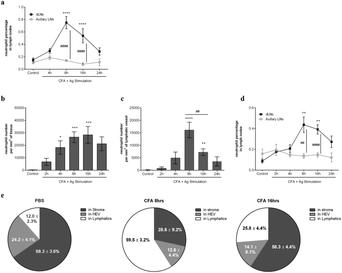
Figure 2. Neutrophil rapidly migrates into lymphatic system of the cremaster muscle during antigen sensitisation in vivo.
Neutrophil migration into the lymphatic system was induced in WT animals following antigen sensitisation with complete Freund’s adjuvant (CFA+Ag). (a) Time course of neutrophil migration into draining LNs (inguinal) or non-draining LNs (axillary) of mice injected intradermally with CFA+Ag and as analysed by flow cytometry. (b) Time course of CFA+Ag-induced neutrophil extravasation in mice injected intra-scrotally with CFA+Ag as visualised by confocal microscopy. (c) Time course of CFA+Ag-induced neutrophil intravasation into the cremaster lymphatic vessels of WT mice as visualised by confocal microscopy. (d) Time course of neutrophil migration into draining and non-draining LNs in mice as analysed by flow cytometry. (e) Quantification of neutrophil localisation in the dLNs of mice stimulated intra-scrotally with CFA (8 or 16 hrs) or with PBS (control), and as analysed by confocal microscopy. Data are represented as percentages of neutrophils present in the HEV, LYVE-1+ vessels and in the stroma of the LNs. Data are expressed as mean ± SEM of N = 5–12 animals (~10 images per cremasters for confocal microscopy) per group from at least 5–10 experiments. Statistically significant differences between stimulated and control groups or between WT and TNFRdbKO mice are indicated by asterisks: *P < 0.05; **P < 0.01; ***P < 0.001; ****P < 0.0001. Significant differences between other groups are indicated by hash symbols: ##P < 0.01; ####P < 0.0001.

