
Figure 1.
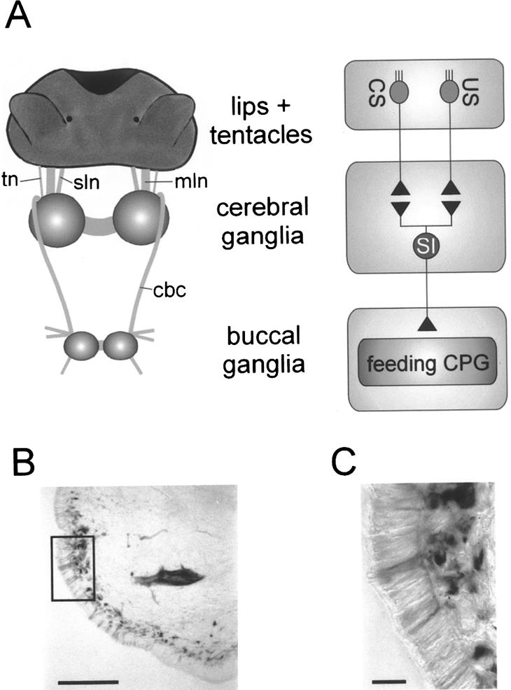
Organization of lip and tentacle sensory pathways to the CNS. (A) Schematic diagram of anatomical organization of lip and tentacle sensory pathways. The lips and tentacles are connected to the cerebral ganglia via the median lip nerve (mln), superior lip nerve (sln), and tentacle nerve (tn). The cerebral-buccal connective (cbc) forms the only connection between the cerebral and buccal ganglia that contain the feeding CPG. Primary chemosensory cells in the lips and tentacles extend direct projections in the lip and tentacle nerves that synapse onto sensory integrating neurons (SI) in the cerebral ganglia (for simplicity, interactions are shown as direct connections). These neurons affect the feeding CPG in the buccal ganglia via the cerebral-buccal connective. (B) Photo-micrograph of putative primary chemosensory cells labeled by backfilling the median lip nerve in a lip section. The picture shows biocytin-labeled neuronal cell bodies arranged in a subepithelial layer. Scale bar, 100 μm. (C) Enlargement of area marked by square in B. Note the fine processes originating from the labeled subepithelial cell bodies that project through the lip epithelium. Scale bar, 20 μm.