
Figure 3.
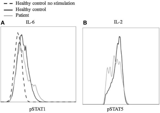
Phosphorylation of STAT1 and STAT5 in response to stimulus. Peripheral blood mononuclear cells from the patient and a healthy control were stimulated with IL-6 (25 ng/mL) (A) and IL-2 (200 ng/mL) (B) for 20 min. The cells were fixed using 4% paraformaldehyde and permeabilized with methanol, then stained with antibody and analyzed by flow cytometry. Cells were gated on CD4+ (A) or CD4+, pSTAT5+ (B) cells. Dashed line shows unstimulated cells from a healthy control. Experiments were repeated at least twice with at least two healthy controls for each experiment.