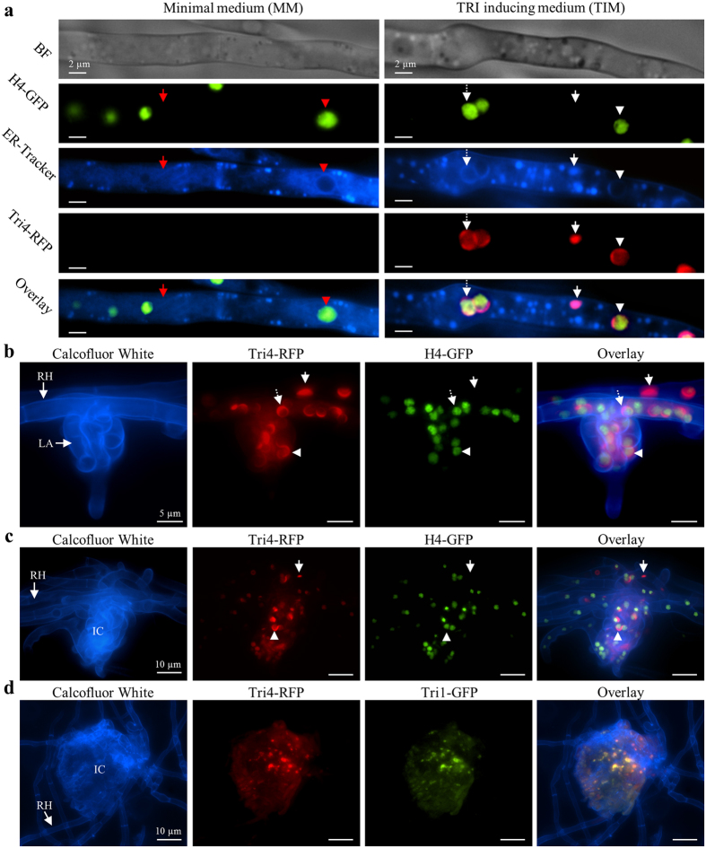
Figure 5. Localization the ER and nuclei in vitro (a) and in planta (b–d).
(a–c) Nuclei and Tri4 visualized with strain H4-GFP/Tri4-RFP in vitro (a, see also Supplementary Fig. S6) and in plant infection structures including lobate appressoria (b) and infection cushions (c and d). (a) Fluorescence of H4-GFP and ER-Tracker are visible in both MM (left column) and TIM (right column). In MM, thin circular structures (red arrowhead) visible by ER-Tracker surround nuclei and thus are perinuclear ER. Reticulate strings (red arrow) not associated with nuclei indicate peripheral ER. In TIM, spheres (dashed white arrow) and crescents (white arrowhead) are visible by ER-Tracker and Tri4-RFP fluorescence and circumscribe nuclei, indicating modified perinuclear ER. Ovoid structures (white arrows) are not spatially associated with nuclei, indicating modified peripheral ER. (b and c) Infection structures of H4-GFP/Tri4-RFP on a palea of wheat 9 days post inoculation stained with Calcofluor White (blue). (b) Runner hyphae (RH) and a lobate appressorium (LA) show crescent structures (arrowheads) and spheres (dashed arrow) by Tri4-RFP, which surround nuclei (green), while ovoid structures (arrows) are not associated to nuclei. (c) Observations similar to those described in b for an infection cushion (IC). (d) The TRI pathway enzymes Tri4 and Tri1 of a Tri4-RFP/Tri1-GFP strain co-localize at modified ER in an infection cushion 9 days post inoculation.

