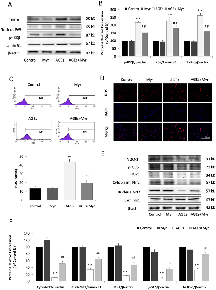
Figure 2. Myr inhibited AGEs-induced inflammation and oxidative stress.
(A) The expression of NF-κB signaling and TNF-α in H9c2 cells by immunoblotting analysis. (B) The relative expression levels of p-IKKβ, nuclear P65, and TNF-α in relation to β-actin were expressed in the bar graphs. (C) Intracellular ROS levels in H9c2 cardiomyocytes evaluated using a flow cytometer. (D) Representive images of ROS staining. Cells with red influorescence indicated elevated intracellular ROS level. (E) Immunoblotting analysis of Nrf2-mediated anti-oxidative enzymes in H9c2 cells. (F) The relative protein expression of Cyto-Nrf2, Nucl-Nrf2, HO-1, γ-GCS, and NQO-1 to β-actin were expressed in the bar graphs. n = 3, **p < 0.01 vs the control group; ##p < 0.01 vs AGEs group.

