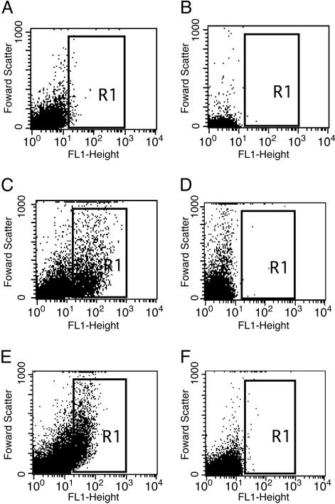
Fig. 5.
Flow cytometric analysis of Pamy-directed GFP expression. S. gordonii (pPamy-′gfp) and V. atypica as monospecies cultures (A and B, respectively) and coculture (C). No increase in fluorescence was seen when S. gordonii containing the gfp reporter plasmid with no promoter, pPE1010, was incubated with V. atypica (D). (E and F) Fluorescence from S. gordonii (pPamy-′gfp) when separated by dialysis tubing from V. atypica (E) and sterile medium (F). Fluorescence (FL1-height) is graphed logarithmically on the x axis. Forward scatter (y axis) is indicative of particle size.

