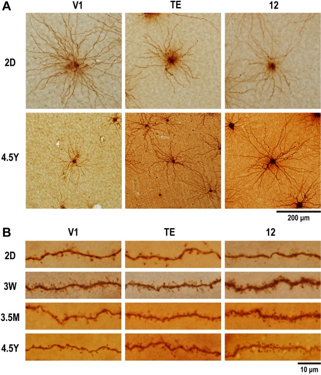
Figure 2.

Photomicrographs of layer-V pyramidal cells individually injected in tangential sections with Lucifer Yellow and reacted for DAB product. (A) Labeled cells for area V1, area TE, and area 12 at 2 days (2D) and 4.5 years (4.5Y). Dendritic branches of pyramidal cell at 4.5Y was smaller than those at 2D in V1, did not change in area TE, and became greater at 4.5Y than at 2D in area 12. (B) Dendritic spines along a dendritic segment in the three areas at each of the four time points. Dendritic segments were poor in spines at 2D, most spinous at 3W in V1, at 3W to 3.5M in area TE and area 12, then spines were pruned toward adulthood.
