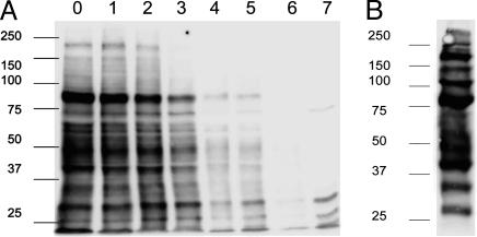
Fig. 1.
Multiple proteins transfer from NK to target cells during inhibitory interactions. (A) YTS-TG was biotinylated and subsequently coincubated with 221/Cw6 target cells for 1 h, after which target and effector cells were separated by FACS. At various times after separation from NK cells, target cell lysates were analyzed by Western blot with streptavidin-HRP. Lanes 0–6 correspond to days 0–6 after coculture and lane 7 is a control target-cell lysate. (B) Biotinylated YTS-TG cell lysates were analyzed by Western blot with streptavidin-HRP. Data shown are representative of four independent experiments. Numbers at left indicate molecular mass (kDa).

