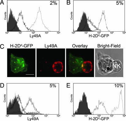
Fig. 4.
Bidirectional transfer of proteins across the inhibitory murine NK cell IS. Sorted Ly49A+ LAK NK cells were coincubated with EL4-Dd-GFP cells, stained with anti-Ly49A mAb, and analyzed by flow cytometry. (A) EL4-Dd-GFP target cells acquired Ly49A (black line) from LAK NK cells (gray line) compared with control noncoincubated target cells (filled histogram). (B) LAK NK cells acquired H-2Dd-GFP (black line) from EL4-Dd-GFP target cells (gray line) compared with LAK NK cells alone (filled histogram). (C) A 3D reconstruction of fluorescence in a conjugate between a Ly49A+ NK cell and EL4-Dd-GFP stained for Ly49A. (Scale bar, 4 μm.) (D) EL4-Dd-GFP cells acquired Ly49A (black line) from Ly49A+ splenocytes (gray line) in comparison to target cells which had not been cocultured (filled histogram). (E) Naive NK cells acquired H-2Dd-GFP (black line) from the target cells (gray line) compared with noncoincubated NK cells (filled histogram). Numbers in histograms represent the percentage acquired.

