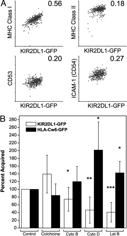
Fig. 5.
Factors influencing the amount of KIR2DL1-GFP transfer to target cells. (A) Conjugates of YTS-TG- and PKH-26-labeled 221/Cw6 cells were isolated by FACS, disrupted with EDTA, and analyzed for expression of MHC class I, MHC class II, CD53, or CD54. Dot plots show the level of expression of MHC class I, MHC class II, CD53, and CD54 on PKH-26+ target cells plotted against the acquired KIR2DL1-GFP fluorescence. Results are representative of three independent experiments. Numbers in the top right corner of each dot plot represent the slope of the linear regression line fitted to the data points. (B) Treated target cells were coincubated with NK cells and analyzed by flow cytometry to assess changes in the amount of KIR2DL1-GFP transferred to target cells (white bars), or the effect on transfer of HLA-Cw6-GFP to NK cells (black bars). Cyto, cytochalasin; Lat B, latrunculin B. Results are shown as a percentage of acquisition in untreated samples. The mean and error bars for nine experiments are shown. Paired Student's t tests were performed. *, P < 0.05; **, P < 0.01; ***, P < 0.001.

